RFLP / MstII test for Sickle-Cell Anemia
RFLP / MstII test for Sickle-Cell Anemia
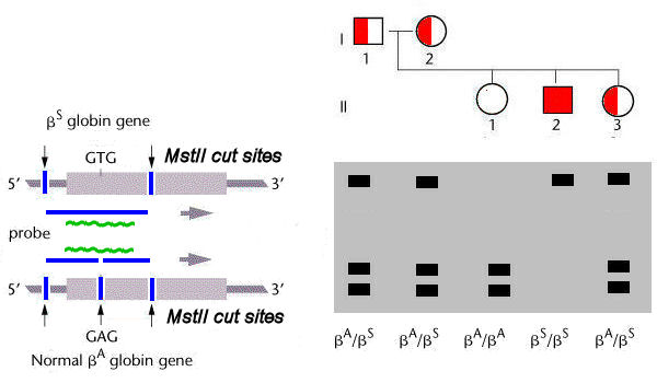
Sickle-cell Anemia is a
molecular disease caused by a mutation in the beta-globin
gene. The difference between the standard ![]() T) in the second position of the sixth codon
of this gene. The sequence of the standard A
allele (CCTGAGG) happens to correspond to an
MstII restriction site
(CCTNAGG), which is altered in the
T) in the second position of the sixth codon
of this gene. The sequence of the standard A
allele (CCTGAGG) happens to correspond to an
MstII restriction site
(CCTNAGG), which is altered in the
In the genetic
test for the S
allele, total DNA from
the individual tested is digested with MstII and run in a Southern Blot. The
blot is hybridized with a probe
specific for the beta-globin
gene. If the standard
Important:
this particular test depends on the coincidence that the
nucleotide substitution responsible for the sickle-cell allele
happens to occur in such a way as to create an RFLP: the absence of the MstII site does not itself cause sickle-cell
anemia, but is instead a genetic
marker for the allele.
Homework:
Suppose this experiment were done by amplifying the
beta-globin gene by PCR, then cutting the product with
MstII
and separating them by electrophoresis as above. How many bands would be
expected in the heterozygote? Explain. Draw
the expected result.