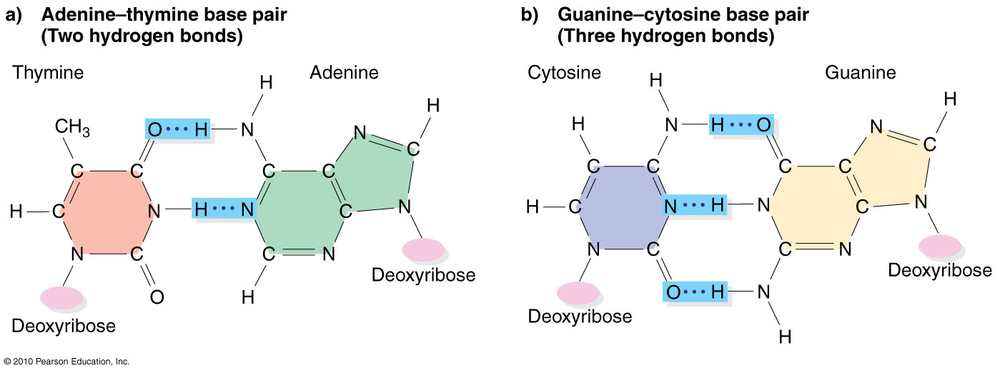
A+T and G+C base pairs have similar structures
The combination of a two-ring purine (A or G) and a single-ring pyrimidine (T or C) forms a base pair (A+T or G+C ) with three
co-planar rings. The distance between the backbones and the
orientation of the bonds to the deoxyribose are essentially
identical for either pair.
The A+T pair is held together by two H-bonds versus the G+C with three. A convenient mnemonic is that
"A" has two "toes" paired to the two
"ears" of the "T". G and C are
rounded: "G" has the two ends in "C",
plus the crossbar, for three bonds.
The bases are often represented as
different colors, according to conventions adopted for
computer visualization in the first generation of automated
DNA sequencer. In this scheme, A C
G T . The yellow G
is easy to read against a black background on a
sequencer computer screen,
but when written on paper is represented by a black G.
Rendered this way, the AT pair shows "Christmas"
colors, and the GC pairs shows "Easter"
colors.
[It was not appreciated for a number
of years that this color scheme made life difficult for
red-green color-blind male scientists. Modern
machines offer a choice of visualization colors that
avoid this problem].