Roux's
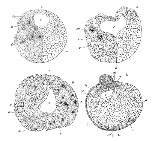
experiment on frog eggs (1888)
Roux
pierced one cell [here, the right side] of a two-cell frog embryo with
a hot needle. This cell continued to divide through the blastula stage [top
row: large cells on the right, while the other [left] side
continued to develop essentially normally. The blastocoel
(F) is apparent in both pictures. A sagittal
section through the left side (below, left] shows normal cell
movement expected for a gastrula,
including epiboly of the dorsal lip (Ee)
inward, giving rise to the production of the three cell
layers. However, subsequent development produced only the left
side of a frog, as shown by a cross-section through the
embryo, where neurulation
has produced the folds of the neural plate (Md) on the
left side, and the right side remains undifferentiated.
Roux concluded that development
was mosaic, and that
already at the two-cell stage determinants
in the single fertilized eggs determined
left- vs right-side development. Subsequent divisions
further divided determinants so as to determine the fate of
the daughter cells. Thus embryonic development is a
consequence of factors internal to
the embryo being partitioned and gradually manifesting
themselves.
Roux
operated without knowledge of Mendelian Genetics, and
neglected the possibility that presence of the 'killed'
right-hand cell might affect development of the un-operated
left side. His results contrast with those of Hans Driesch's
experiment.