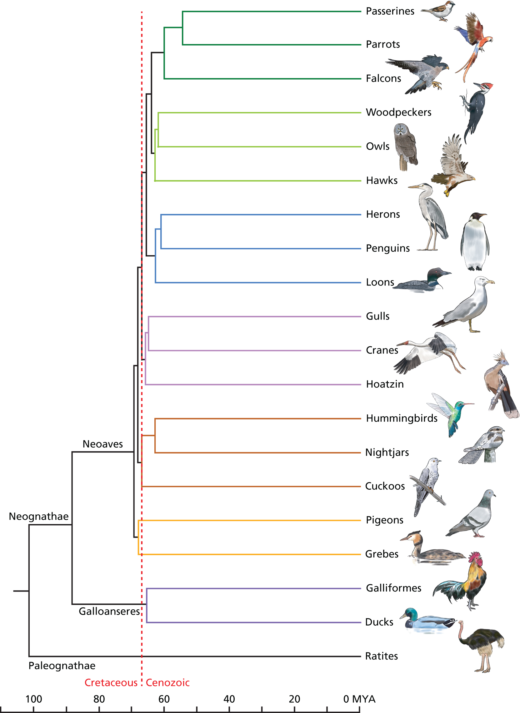
A modern interpretation of bird evolution, based on
molecular data
(ED Jarvis et
al. 2014. Science, 346:1320)
The historical
understanding of the evolutionary origin of taxonomic orders of
moderns birds was based on a fossil record that identified
bird-like forms as far back as 100 MYBP. In particular, the
occurrence of flightless Ratite forms on the three
continents of Gondwanaland (Ostriches in
Africa, Rheas in South America, and Emus in
Australia) suggested a common origin before the breakup of the
super-continent. Given this timing, early molecular studies of
birds then suggested that the "Molecular
Clock" in birds ran unaccountably "slow",
in contrast to mammals and other vertebrates classes. That is,
for example, the molecular differences between Galliformes (Chickens
and relatives) from Anseriformes (Ducks and relatives)
was substantially less than for a pair of mammalian
orders that had diverged 100 MYBP. The seeming rate
inconsistency of the Molecular Clock was used as an argument
against its general use.
Modern studies of
the molecular evolution of bird orders shows instead that the
Ratites separated from other bird orders long before the general
Adaptive
Radiation of modern bird orders (Neoaves &
Galloanseres) 65 MYBP. Note that, even though the
ancestors of chickens and ducks had already separated from other
birds 90 MYBP, the divergence of these two orders occurs
simultaneously with the main radiation. This suggests the
hypothesis that the sudden diversity of birds was coincident
with the disappearance of the (other) Archosaur orders
at the K-T (Cretaceous - Tertiary) Boundary,
possibly due to the clearing of old adaptive niches and creation
of new ones. The adaptive
radiation of Mammalia also occurred at this time,
probably under similar circumstances.
This example
demonstrates the success of molecular data in establishing a
reliable, dated phylogenetic history for groups of organisms.
This timescale can then be used to explain their morphological
evolution, rather than the classical tail-chasing process of
using morphology to create a phylogenetic scheme,
and then using that scheme to explain morphology.