RNA Translation: RNA makes Protein
In principle:
Translation of messenger
RNA (mRNA) takes place on ribosomes,
which include ribosomal RNA (rRNA),
with the help of transfer RNA (tRNA)
Structure of rRNA &
tRNA
ribosomal RNA (rRNA)
rRNA + ribosomal protein  ribosomes [iG1
6.09]
ribosomes [iG1
6.09]
Structure of rRNA: stems
& loops [iG1 6.05]
stems: double-stranded (dsRNA)
loops:
single-stranded (ssRNA)
Structure of eukaryotic ribosomes [iG1
6.04] [iG1 6.14b]
Large Subunit (LSU) = 60S = 28S rRNA
+ 5.8S + 5S rRNA + 50 proteins
Small Subunit (SSU) = 40S = 18S rRNA
+ 33 proteins
= 80S
monosome
A site (Aminoacyl), P site (Peptidyl), & E site (Exit) [APE complex]
transfer RNA
(tRNA)
the adaptor molecule: ~30 tRNA types
2-dimensional 'cloverleaf' model
[iG1
6.10]
small: 75 ~ 90 nucs
stems & loops
D-loop & T C-loop ( = pseudo-uridylic
acid)
C-loop ( = pseudo-uridylic
acid)
tRNA
characterized by 2o
modified bases [iG1 6.19]
amino-acceptor stem
3' - ~~~~ CACCA - 3'
5' -~~~~ G
- 5'
anticodon loop
specificity of tRNA
for mRNA determined by 3-ribonucleotide sequence
3-dimensional structure is
an "L" [iG1
6.12]
D- & TC-loops fold back on
each other
Charged tRNA: aminoacyl synthetase(x) forms ester linkage between
3'-A of
amino-acceptor stem of tRNA(x) joined to COOH of amino acid(x)
~20
synthetase
types 'recognize' correct
anticodon loop
isoacceptance:
one-to-one correspondence between synthetase & amino
acid
RNA Translation: Protein
Synthesis
Ribosomes "read" mRNA & assemble polypeptide according to the Genetic Code
Initiation at start codon (AUG)
SSU binds at Shine-Delgarno sequence (-6
nucs)
Initiation
Complex consists of mRNA, ribosome,
& tRNA
Multiple
complexes form on a single mRNA: polysome (polyribosome)
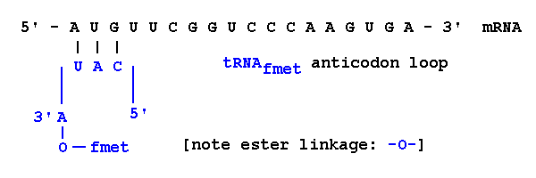
tRNAfmet
always added first [N-formyl-methionine
in prokaryotes]
In
simplified form,
5'-AUG-3' codon in mRNA
|||
3'-UAC-5' anticodon in tRNA
5'-CAU-3' if anticodon is written 5' 3'
3'
Elongation: addition of amino acids according to Genetic Code
Amino acids are joined via peptide bonds (see next section)
Think of mRNA as stationary: ribosome moves
along it 5'3'
peptidyl (P)
site on 5'
end,
aminoacyl (A)
site on 3'
end
first AUG codon [for met] is in P site
second UUC codon enters A site
corresponding tRNAphe
enters A site
peptide
bond formed between fmet
& phe
P site amino acid
transferred to A site
amino acid
uncharged
tRNA released from P
site (passes to E site)
amino end of fmet remains unchanged ]
and so
on ...
growing polypeptide in P site joins single amino
acid in A site
"Wobble": pairing of codon / anticodon
goes 5'3'
on codon [iG1
7.25, 26]
last position can miss-pair with either purine / pyrimidine
Fewer tRNA species needed:
Ex.: three tRNAser species for six codons
| tRNA Anti-codon |
Alternative Serine
mRNA codons |
| 3'- AG G -5' | 5'- UC C / U -3' |
| 3'- AG U -5' | 5'- UC A / G -3' |
| 3'- UC G -5' | 5'- AG C / U -3' |
Termination: release of polypeptide
mRNA + tRNA(aan-...-aa3-aa2-aa1 )
here: mRNA + tRNA(lys-pro-gly-phe-fmet)
stop codon (UAG, UAA, or UGA) enters A site
no corresponding tRNA:
release factor
cleaves polypeptide from terminal tRNAn
polypeptide product is: lys
- pro - gly - phe - fmet
A talkie animation
of transcription & protein synthesis
Griffiths et al.
(1996) Fig. 13-7
is a nice summary (HOMEWORK
#11)
Bioinformatics of DNA, mRNA, & Protein
5'- G T A A T C C T C - 3' DNA sense strand
5'- G U A A U C C U C - 3' mRNA
N - val - ile - leu - C protein
This is a logical,
not a biochemical, relationship:
Because mRNA is transcribed from the template strand,
it "looks like" the sense strand (except for
'U').
The information
content of the DNA sense
strand and mRNA are identical
Protein sequences can be read directly from DNA:
Read the sense strand
in the 5'3' direction,
Substitute 'T' for 'U' in the code table [or in
your head]
Computer programs (Chromas, Sequencher,
etc.) do this automatically
There are three reading frames
on either strand
X
two 5'3'
strands ![]() six possible ways to
read dsDNA
six possible ways to
read dsDNA
Open
Reading Frames suggest protein sequences
Deducing protein sequences from random DNA
sequences is a major research
activity
Bioinformatics:
extraction of information from large macromolecular datasets
The following clues are useful:
Remember that all prokaryotic coding
sequences:
are read only in the 5'3' direction
begin with a "start" (AUG)
codon
end with a "stop" (UAG, UAA, or
UGA) codon.
Ex.: a typical exam problem
is to identify a polypeptide
of six amino acids from a dsDNA molecule
But: in real life research,
cloned eukaryotic DNA
may not have start or the stop codon for a complete
protein,
[and
not all AUG codons
are 'start' codons]
and may be include an intron with one or more 'stop' triplets .
Do not assume that a dsDNA
molecule is read from left to right, on the top strand
Homework #12:
Practice DNA
"Translation" problems [PDF download
version]
There's
an App for that: RandORF
for 'translation' problems
All text material © 2016 by Steven M. Carr