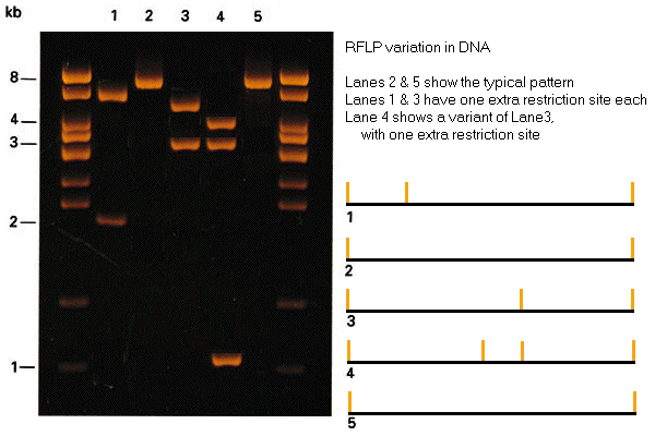
Gel
electrophoretic detection of RFLP variation
SNPs (single-nucleotide polymorphisms)
that create or destroy restriction sites lead to RFLPs (restriction fragment length
polymorphisms) detectable by gel electrophoresis. The
outside lanes are molecular size standards. Lanes 2 &
5 show the typical pattern of a single 8 Kbp fragment.
Lanes 1 & 3 show two variants that result from the presence of
either of two additional restriction sites. Lane 4 shows a variant
of the genotype in Lane 3, with an extra restriction site.
The restriction maps shown at right cannot be
reconstructed from the gel data shown, but could be mapped with
reference to data from other restriction enzymes.