
Molecular Biology of Hemoglobin in
Sickle-Cell Anemia
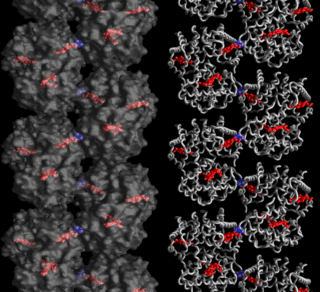
The first
figure shows a sickle-cell hemoglobin fiber, with heme groups in red and the valine residue
in blue. The fiber consists of two strands: note that the valine residues
make intermolecular contacts between the two strands,so as to
stabilize the structure.
The second figure is a close-up stereo diagram of the contact: the regions labelled E & F are in the beta-globin subunit of a hemoglobin one strand, and the A & H regions are in the other fiber. The yellow Val residue on the right-hand molecule fits into a pocket formed by amino acids 84, 85, 87, & 88 in the left hand molecule.
[To view the stereo figure: gaze at a point between the two
drawings and let your eyes cross; blink to superimpose the two
out-of-focus images into one in-focus image].
Figures © 2009 by W. Royer; text material © 2014 by Steven M. Carr