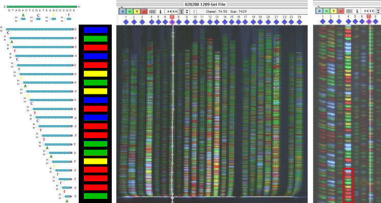
Gel images from an automated DNA sequencer
Gel images from an automated DNA sequencer
From the original
DNA template, the dideoxy sequencing reaction synthesizes
a new DNA strand that incorporates a set of
fluorescently-labeled ddNTPs into the DNA (left).
The newly synthesized DNA is separated by size in an
electrophoretic field at high voltage, and the fluorescence
colours are activated by a scanning laser, tracked, and
read by a photometer. The fluorescent dyes used are four
slightly different wavelengths of green:
the gel image (middle) is a "false colour" representation, in
which these wavelengths are shown as distinct A C G & T. Each of 24 different
DNA sequences is a separate sequencing reaction;
automatic tracking for one lane is shown by the white
trace. In the magnified view (right), the sequencing
ladder in each lane has four different coloured bands that
correspond to each of four DNA bases. Each channel
is converted to an individual chromatogram for that DNA
sequence; the order of coloured peaks is converted to a DNA sequence. For example,
the first few bases of the fourth lane boxed in red will be read
as ATTTGAATTC
.
Because the four
bases appears as different colors, all four reactions for any
sample can be run in and read in a single lane, rather than four
lanes when a 32P label is used.
Automated DNA sequencing revolutionized the field and
made possible the Human Genome Project. The 24 reactions
show here cover the complete 16 Kbp sequence of a human
mtDNA genome. The most advanced machine of that time could
run 96 samples simultaneously.
HOMEWORK: Popular accounts like the movie "Jurassic Park" suggest that "DNA sequences are read by a laser'" Explain why this is incorrect.